
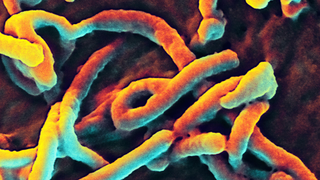
The NIAID (National Institute of Allergy and Infection Diseases) has shared these microscopic images of Ebola on the organization’s Flickr account. The scanning electron micrographs include an image of the virus budding from the surface of a Vero cell of an African green monkey’s kidney epithelial cell line. The institute is working on developing Ebola vaccines, including one that will prevent the transmission of the virus from animals to humans.
Speaking to Fox News amidst a “close the borders” panic yesterday, NIAID director Anthony Fauci reiterated that a quarantine to this effect will not help the relief and containment efforts. He also stated that using Ebola as a weapon of bioterror is “not the most effective” and nature is “worse than a bioterrorist.”
Meanwhile, New York City health official are optimizing their preparedness in the possible case of outbreak in the city, a week since one case of Ebola has been reported in Dallas. The initiative includes new protocols, like asking all people reporting symptoms of vomiting and fever if they had been to West Africa in the last three weeks. Dr. Irwin Redlener, the director of the National Center for Disaster Preparedness at Columbia University and a special adviser to Mayor Bill de Blasio, emphasized that “New York’s West Africans know they can seek medical care regardless of their immigration status or ability to pay.” (Images: NIAID)
