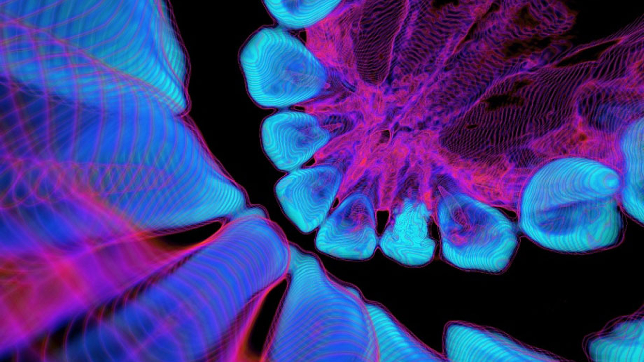
No, these aren’t those way-trippy black light dorm posters they sell at Spencer’s Gifts. They’re actually software-enhanced medical images of teeth, rotting teeth, the inside of a left nostril, heart valves, the brain’s fourth ventricle, stress lines in the skull, and cranial blood vessels; respectively. They just happen to be really, really, mind-meltingly psychedelic.
Hong-Kong based radiologist Dr. Kai-hung Fung had no intention of producing “art” when he generated these images. The project came about purely by accident when he was asked to generate complex 3-D anatomical images for surgeons to visually prepare themselves before operating. He stacked up CT scans of different organ “slices” shot from different angles, indicating changes in depth by using different colors and contour lines.
According to Slate, “Dr. Fung’s ‘4-D visualizations’ (short 3-D videos) aid surgeons by ‘showing changing perspectives and relative relationships of various anatomical structures … [matching] the surgical or endoscopic field of view.'”. Don’t you love when things that are sweet to stare at also benefit society?
Dr. Fung is currently working on producing 3-D CT images of flowers and biological specimens.


